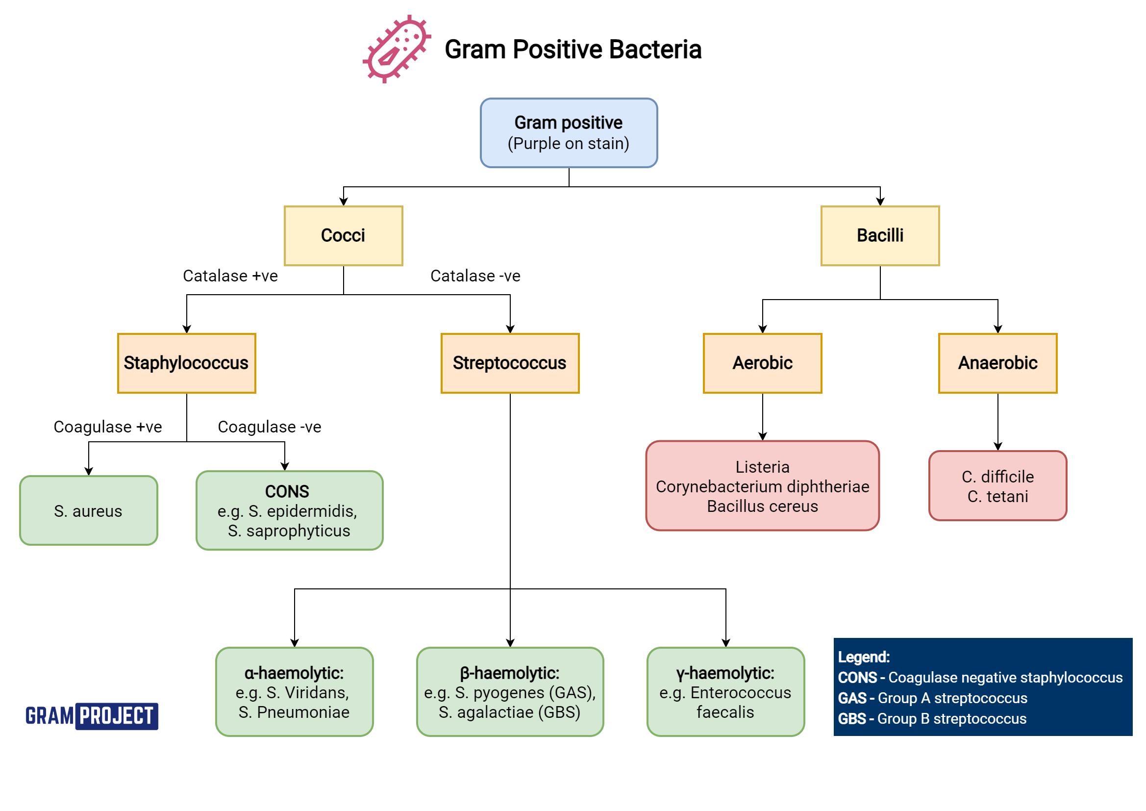
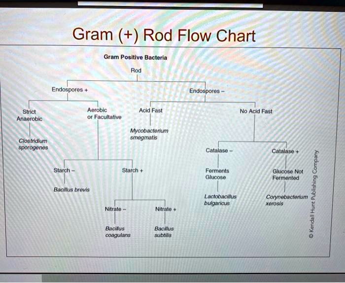
Gram Positive Flow Chart
Gram Positive Flow Chart - Bacillus, clostridium, corynebacterium, listeria, and gardnerella. Lugdunensis found in abscesses and serious wounds; Web identify different types of bacterial morphology seen on a gram stain. Gram staining is one way scientists can. How agar media helps us identify microbes. Move your mouse over an item on the graphic and if your arrow turns into a hand click on it and you will go to another place in the notebook.) click on gram positives to determine how and when to perform the tests indicated above. Web what are they? Web aerobic gram positive cocci flowchart. In this study, a total of 124 strains, including 13. Bacteria are identified in medical laboratories by various methods, identification of microbes. Gram positive (g+), catalase positive (+) cocci. Use flowcharts and identification charts to identify some common aerobic gram positive microorganisms. The bacterial cell wall of these organisms have thick peptidoglycan layers, which take up the purple/violet stain. Web after your bacteria have been isolated and you have good results from your gram stain, begin to follow the flow chart below.. (the graphic below is clickable. Web gram positive flow chart. Web identification of negative onpg enterobacteriaceae. That exists almost exclusively within host cells, i.e., intracellular bacteria (e.g., chlamydia) Coagulase negative staphylococci in multiple blood cultures. Web what are they? Web gram positive cocci obligate anaerobic peptostreptococcus spp., peptinophilus spp., parvimonas spp., anaerococcus spp., atopobium spp., f. Flow chart of gram positive organisms created date: A gram stain test, which involves a chemical dye, stains the bacterium’s cell wall purple. Gram positive (g+), catalase positive (+) cocci. Web this paper reviewed core concepts of interpreting bacterial culture results, including timing of cultures, common culture sites, potential for contamination, interpreting the gram stain, role of rapid diagnostic tests, conventional antibiotic susceptibility testing, and automated testing. Web identify different types of bacterial morphology seen on a gram stain. Bacillus, clostridium, corynebacterium, listeria, and gardnerella. Associate various biochemical tests with. Web identify different types of bacterial morphology seen on a gram stain. Web aerobic gram positive rods flowchart. Web identification of negative onpg enterobacteriaceae. Move your mouse over an item on the graphic and if your arrow turns into a hand click on it and you will go to another place in the notebook.) click on gram positives to determine. In this study, a total of 124 strains, including 13. Identification of bacteria, identification algorithm gram positive bacillus, identification of gram positive cocci, identification of. The bacterial cell wall of these organisms have thick peptidoglycan layers, which take up the purple/violet stain. Web aerobic gram positive rods flowchart. Nearly all clinically important bacteria can be visualized using the gram staining. That exists almost exclusively within host cells, i.e., intracellular bacteria (e.g., chlamydia) Web this paper reviewed core concepts of interpreting bacterial culture results, including timing of cultures, common culture sites, potential for contamination, interpreting the gram stain, role of rapid diagnostic tests, conventional antibiotic susceptibility testing, and automated testing. Use flowcharts and identification charts to identify some common aerobic gram. Web aerobic gram positive rods flowchart. Associate various biochemical tests with their correct applications. The gram stain, is a laboratory staining technique that distinguishes between two groups of bacteria that have differences in the structure of their cell walls. Bacteria are identified in medical laboratories by various methods, identification of microbes. How agar media helps us identify microbes. (the graphic below is clickable. The gram stain, is a laboratory staining technique that distinguishes between two groups of bacteria that have differences in the structure of their cell walls. Web identification of negative onpg enterobacteriaceae. Web the gram stain differentiates organisms by the way the react with colored stains: A gram stain test, which involves a chemical dye, stains. (the graphic below is clickable. Web what are they? Web identification of negative onpg enterobacteriaceae. Gram positive (g+), catalase positive (+) cocci. Web gram positive flow chart. Web aerobic gram positive cocci flowchart. How agar media helps us identify microbes. Web gram positive cocci obligate anaerobic peptostreptococcus spp., peptinophilus spp., parvimonas spp., anaerococcus spp., atopobium spp., f. Gram staining is one way scientists can. The bacterial cell wall of these organisms have thick peptidoglycan layers, which take up the purple/violet stain. Lugdunensis found in abscesses and serious wounds; Web this paper reviewed core concepts of interpreting bacterial culture results, including timing of cultures, common culture sites, potential for contamination, interpreting the gram stain, role of rapid diagnostic tests, conventional antibiotic susceptibility testing, and automated testing. Where bacteria can be gram stained the cell shape and clustering are of practical value in identification. Identification of bacteria, identification algorithm gram positive bacillus, identification of gram positive cocci, identification of. Use flowcharts and identification charts to identify some common aerobic gram positive microorganisms. A gram stain test, which involves a chemical dye, stains the bacterium’s cell wall purple. Web after your bacteria have been isolated and you have good results from your gram stain, begin to follow the flow chart below. In this study, a total of 124 strains, including 13. Web identification of negative onpg enterobacteriaceae. (the graphic below is clickable. Move your mouse over an item on the graphic and if your arrow turns into a hand click on it and you will go to another place in the notebook.) click on gram positives to determine how and when to perform the tests indicated above.
gram negative identification flow chart Gram positive Bacteria Flow

Gram Positive Cocci Flow Chart

Gram Positive Organisms Chart

Bacteria Identification Flow Chart Gram Positive Bacteria Flow Chart

Gram Positive Bacteria Flow Chart Diagram Quizlet

Pinterest • The world’s catalog of ideas

Listeria monocytogenes USMLE Strike

Gram Positive Flow Chart *Science and WoNdEr* Pinterest Pharmacy

Microbiology Gram Positive Cocci Flow Chart Gram Positive Cocci

Gram Positive Bacteria Flow Chart Diagram Quizlet
Bacterial Comes In Different Shapes And Sizes ,But One Way Scientist Use To Identify Bacterial Is By Using Gram Stain Test ,Where By Gram Positive Bacterial Appear Purple In The.
Web What Are They?
Bacteria Are Identified In Medical Laboratories By Various Methods, Identification Of Microbes.
The Gram Stain, Is A Laboratory Staining Technique That Distinguishes Between Two Groups Of Bacteria That Have Differences In The Structure Of Their Cell Walls.
Related Post: