Spine Chart With Nerves
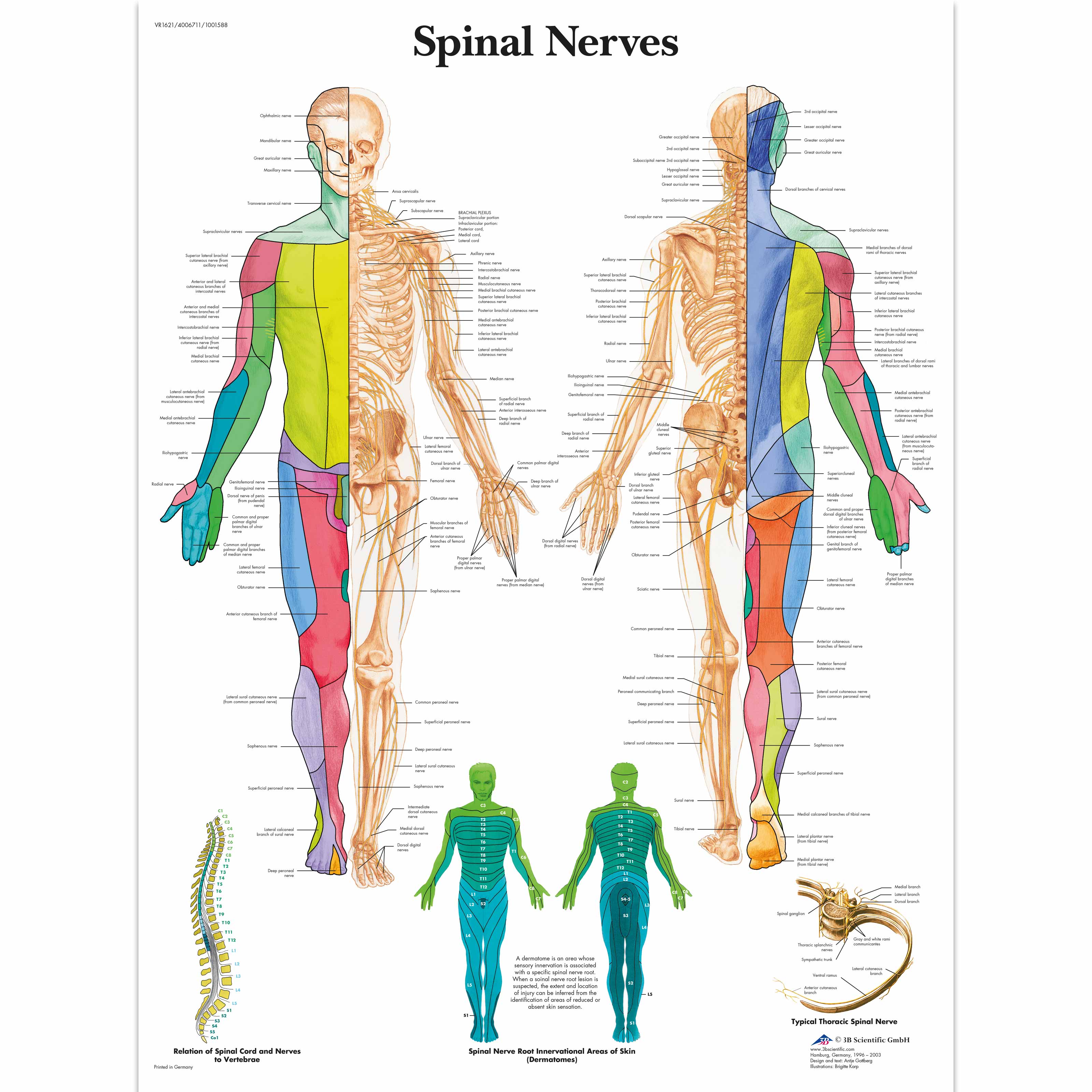
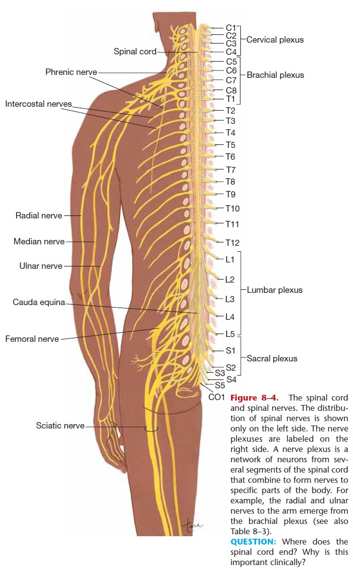
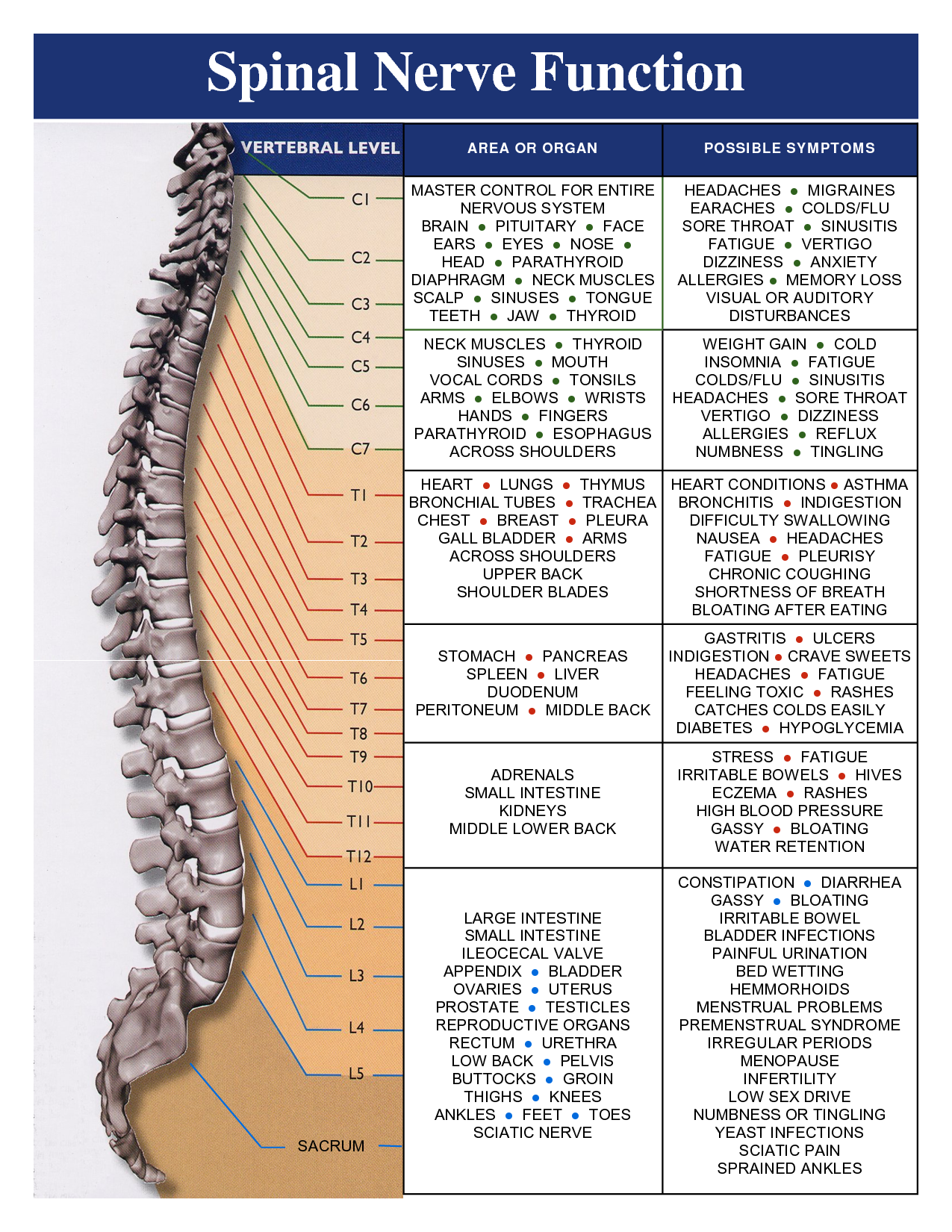
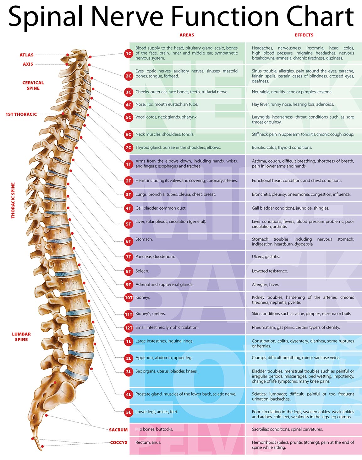
Spine Chart With Nerves - Twelve thoracic spinal nerves in each side of the body called t1 through t12; The back functions are many, such as to house and protect the spinal cord, hold the body and head upright, and adjust the movements of the upper and lower. Web the spinal cord and peripheral nerves. The central illustration shows a posterior view of the spinal nerves exiting from the vertebral column and running throughout the body. The back is the body region between the neck and the gluteal regions. Web the 30 dermatomes explained and located. The spinal cord begins at the base of the brain and extends into the pelvis. The peripheral nerves are responsible for sensations and muscle movements. Web the nerves connected to the spinal cord are the spinal nerves. It comprises the vertebral column (spine) and two compartments of back muscles; What exactly is the spine? The point at which a nerve exits the spinal cord is called a nerve root. Five sacral spinal nerves in each side called s1 through s5; These nerves exit the intervertebral foramina below the corresponding vertebra. The sensory axons enter the spinal cord as the dorsal nerve root. One coccygeal nerve on each side called co1 Your lumbar spine consists of the five bones (vertebra) in your lower back. Web what is the lumbar spine? On the chart below you will see 4 columns (vertebral level, nerve root, innervation, and possible symptoms). The spinal cord begins at the base of the brain and extends into the pelvis. How to use the spinal nerve chart: The ventral root is the efferent motor root and carries motor information from the brain. Many of the nerves of the. Web there are 31 bilateral pairs of spinal nerves, named from the vertebra they correspond to. Five sacral spinal nerves in each side called s1 through s5; On the chart below you will see 4 columns (vertebral level, nerve root, innervation, and possible symptoms). Each spinal nerve is a mixed nerve, formed from the combination of nerve root fibers from its dorsal and ventral roots. Web there are 31 pairs of spinal nerves located at the cervical, thoracic, lumbar, sacral, and coccygeal levels: Your lumbar vertebrae, known. The spinal cord begins at the base of the brain and extends into the pelvis. The peripheral nerves are responsible for sensations and muscle movements. Many of the nerves of the. Web the 30 dermatomes explained and located. What exactly is the spine? Web a very popular and useful chart, the spinal nerves illustrates the spinal nerves and pathways through the body. The two types of nerve roots on each side include: The central illustration shows a posterior view of the spinal nerves exiting from the vertebral column and running throughout the body. Web spinal nerves are mixed nerves that interact directly with. Web a very popular and useful chart, the spinal nerves illustrates the spinal nerves and pathways through the body. The point at which a nerve exits the spinal cord is called a nerve root. Web what is the lumbar spine? Important skeletal structures are included. The roots connect via interneurons. The spinal cord serves as the central pathway for transmitting sensory and motor signals between the brain and the body through specific. Web there are 31 bilateral pairs of spinal nerves, named from the vertebra they correspond to. The dorsal root is the afferent sensory root and carries sensory information to the brain. Web your spinal cordãs nerves branch out. The dorsal root is the afferent sensory root and carries sensory information to the brain. Web what is the lumbar spine? The central illustration shows a posterior view of the spinal nerves exiting from the vertebral column and running throughout the body. Ventral root (located in front) that carries motor signals from the brain to that nerve root’s myotome, which. Your spine has 33 stacked vertebrae (small bones) that form the spinal canal. The roots connect via interneurons. Web eight cervical spinal nerves on each side of the spine called c1 through c8; Each spinal nerve is a mixed nerve, formed from the combination of nerve root fibers from its dorsal and ventral roots. Each nerve forms from nerve fibers,. For most spinal segments, the nerve roots run through the bony canal, and at each level a pair of nerve roots exits from the spine. The central illustration shows a posterior view of the spinal nerves exiting from the vertebral column and running throughout the body. The back is the body region between the neck and the gluteal regions. Web below is a chart that outlines the main functions of each of the spine nerve roots: A long, tubular bundle of nerves that extends from the brainstem down the vertebral column, protected by fluid in the spinal canal and surrounded by ligaments and bone for protection. The sensory axons enter the spinal cord as the dorsal nerve root. 1 pair of coccygeal nerves (co1). The arrangement of these nerves is much more regular than that of the cranial nerves. Web many nerves come from the spinal cord, pass through foramina (holes) formed by notches of 24 vertebrae in the movable spinal column, and innervate or supply specific areas and parts of the body.2 whenever specific areas or parts of the body are malfunctioning, generalized and/or specific symptoms are possible. The spinal cord serves as the central pathway for transmitting sensory and motor signals between the brain and the body through specific. Ventral root (located in front) that carries motor signals from the brain to that nerve root’s myotome, which is the group of muscles that it controls. Web 8 cervical spinal nerves. All of the spinal nerves are combined sensory and motor axons that separate into two nerve roots. Your lumbar spine consists of the five bones (vertebra) in your lower back. Each nerve forms from nerve fibers, known as fila radicularia, extending from the posterior (dorsal) and anterior (ventral) roots of the spinal cord. Web each level of the cervical spine has four nerve roots—two on each side—that branch off from the spinal cord.
Anatomical Charts and Posters Anatomy Charts Spinal Nerves Paper Chart

36 best images about body system correlation charts on Pinterest

Spinal Nerve Anatomy Diagram

Spinal Nerve Chart

Spinal Nerves Anatomical Chart Spine and Cranial Nervous System

Vital Connections Briggs Chiropractic Clinic

17 bästa bilder om Anatomy & Physiology på Pinterest Människans

Spinal Nerve Chart Spinal nerve, Varicose vein remedy, Spinal

Cervical Spinal Nerve Chart

Where Do Your Nerves Go? ADIO Chiropractic Center
Your Lumbar Vertebrae, Known As L1 To L5, Are The Largest Of Your Entire Spine.
The Ventral Root Is The Efferent Motor Root And Carries Motor Information From The Brain.
For The Most Part, The Spinal Nerves Exit The Vertebral Canal Through The Intervertebral Foramen Below Their Corresponding Vertebra.
Web The Lumbar Spinal Nerves Innervate The Lower Limbs.
Related Post: